|
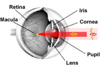
Fig.
1
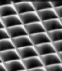
Fig.
2
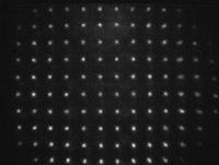
Fig. 3

Fig. 4

Fig. 5
|
It is well known
that the human eye
is not a perfect optical system. The imperfection of the eye
optics is caused by
aberrations induced by
the cornea, lens, and ocular media. Generally, the human eye
without pathologies in refraction (like myopia and astigmatism),
can be considered as almost diffraction limited at the eye pupil
diameter of about 2mm. When the iris is opened wider, the
optical aberrations significantly reduce the vision quality.
There are two major tasks
where precise measurements and correction of eye aberrations is
vital: excimer laser vision correction and clinical diagnostics
of the retina.
Laser vision correction,
like LASIK
with novel "flying spot" lasers, requires precise knowledge
about the overall aberrations of the eye. In such a case, it's
possible to perform "customized ablation" and to significantly
improve the operation success rate, especially in complex cases.
Among several possible
techniques for aberration measurements, the Shack-Hartman
wavefront analyzer gathered worldwide attention. The majority of
custom cornea ablation systems (like VISX, Carl Zeiss Meditec,
Baush&Lomb, etc) utilize Shack-Hartman aberrometers.
The basic principle of
human eye aberration measurement with a conventional
Shack-Hartman wavefront
sensor is illustrated
in Figure 1.
The eye focuses a low-power laser beam on the
retina,
producing a virtual point source. The scattered laser radiation
is reflected back and acquires the aberrations induced by the
ocular media, lens, and cornea. A lenslet array
(Fig. 2)
samples the distorted wavefront, forming a regular spot pattern
called a Hartmanogram
(Fig. 3).
The deviations of the spot centers from the reference grid are
proportional to the local slopes of the wavefront. Thus, by
reconstructing the wavefront slope map with subsequent Zernike
wavefront approximation
[
see Zernike
polynomials
]
it’s possible to obtain the complete aberration map of the eye.
However, in reality the
aberrations of the living eye are not stationary due to many
factors, including tear film variations, fluctuations of
accommodation, etc. Thus, the “single snapshot” type of
measurement does not provide any reliable data for the diagnosis
and treatment. Our group was one of the first who suggested and
implemented the principle of dynamic aberrometry.
In dynamic aberrometry,
the aberrations of the eye are measured with a temporal
resolution exceeding the shortest period of aberration
fluctuations. Since the spectrum of the aberration fluctuations
(Fig.4) is located within the 10-12 Hz bandwidth, the sampling
rate should exceed 20-25Hz. Having a series of dynamic data, one
can (automatically or manually) figure out the most significant
and stationary aberrations, which need to be corrected by
refractive corrections techniques. The poster
[
Utility of dynamic aberrometry for acuity measurements and
testing
]
(in PDF, 1.55 Mb)
contains more detailed information on dynamic aberrometry.
During last few years we
have been developed several aberration diagnostic instruments
capable of measuring human eye aberrations in real time (up
to 80 measurements per second).
The unique feature of the instruments is a scanning reference
spot, which greatly improves the measuring accuracy and speed
(US patent
#US 6331059 B1,
Russian Patent
#2268637).
The instrument has powerful analytical software with open data
exchange capability. A more detailed specification of the
instrument can be found
[
here
].
Another aspect of the
visual optics research carried out by our group concerns
studying the influence of different aberrations on the visual
acuity. For this purpose, in 2002 we (together with Dr.M.Mrochen*
from Swiss National
Institute of Technology)
developed a dynamic aberrometer with adaptive optics. This
instrument (Fig. 5)
includes a deformable
mirror capable of
compensating aberrations up to the 4th order (such as coma,
trefoil, spherical aberration, etc.)
[
see Zernike
polynomials
].
The aberrations of the human eye can be automatically
compensated (or introduced) while the patient looks at a
specific target. Thus, one can study the influence of
aberrations on the subjective visual acuity and simulate the
results of customized refraction correction procedures.
The main field of
application of the instruments is the research in the field of
visual optics, such as aberration dynamics, accommodation, etc.
The technology is currently commercialized by
Visionica Ltd.
A special version of the
aberrometer has been developed for the Russian excimer laser
complex MicroScan 2000.
[
Physics Instrumentation Center
]
|
|
PUBLICATIONS:
Larichev A.V., Ivanov P.V., Iroshnikov N.G.,
Shmalhauzen V.I., Otten L.J., Adaptive system for eye-fundus
imaging, Quantum Electronics, 32, N10, 2002, p.902.

A. Larichev, P. Ivanov, I.
Irochnikov, S.C. Nemeth, A. Edwards, P. Soliz, High Speed
Measurement of Human Eye Aberrations with Shack-Hartman Sensor.
[ARVO Abstract]
Invest Ophthalmol Vis Sci., 42 (2001) 897
A.V.Larichev, P.V.Ivanov,
I.G.Irochnikov, V.I.Shmal'gauzen, Measurement of eye aberrations
in a speckle field, Quantum Electronics, 31 (2001) 1108
Jos j. Roesema, Dirk E.M.
Van Dyck, M.-J. Tassignon, Clinical comparison of 6
aberrometers. Part 1: Technical specifications, J Cataract
Refract Surg 2005; 31:1114–1127
Jos j. Roesema, Dirk E.M.
Van Dyck, Micha Pauw, Bas van Der Spek,
M.-J. Tassignon, Clinical comparison of 5 commercially
available aberrometers II: statistical
comparison on a test group
N. G. Iroshnikov, A. V.
Larichev, Adaptive optics in ophthalmology, Proc. SPIE Vol.
6284, 62840B. Sep 2006
Goncharov A.S.,
Larichev A.V.,
Iroshnikov N.G., Ivanov V.Yu.,
Gorbunov S.A., Modal tomography of
human eye aberrations,
Laser Physics, 2006, V.16, N12, p.1689.
Goncharov A.S.,
Larichev A.V., Speckle Structure of a
Light Field Scattered by Human Eye Retina,
Laser Physics, 2007, V.17, N9, p.1157-1165
|

